Atrial Fibrillation V Fib Ecg


Atrial fibrillation is verified on the ecg resting ecg holter ecg event recorder.
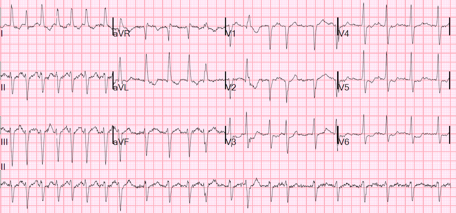
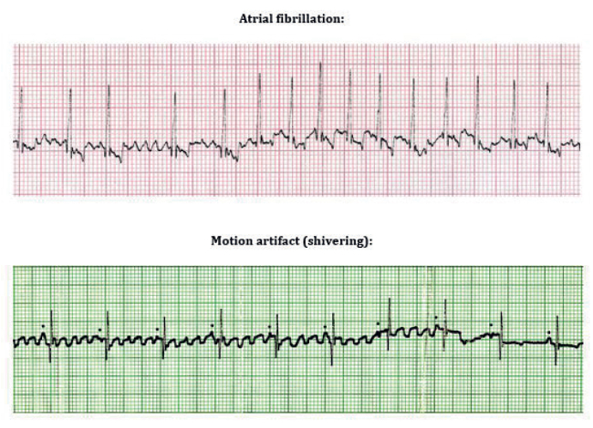
Atrial fibrillation v fib ecg. In atrial flutter there is a sawtooth pattern on an ecg. In a fib you will see many fibrillation beats instead of one p wave. In this atrial fibrillation ecg review the ecg criteria to diagnose atrial fibrillation afib including atrial fibrillation with rvr coarse atrial fibrillation and other af scenarios are.
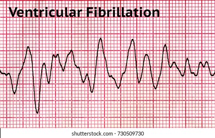
A glitch in the heart s electrical system makes its upper chambers the atria beat so fast they quiver or fibrillate. Ecg features of atrial fibrillation. Atrial fibrillation afib and ventricular fibrillation vfib are both a type of abnormal heart rhythm arrhythmia.
Absence of an isoelectric baseline. If the patient may have coronary heart disease exercise stress test exercise ecg should be considered. This results in the inability of the heart to contract.
Atrial fibrillation or a fib can lead to fatal heart complications if it reaches a severe enough stage. Atrial fibrillation is caused by irregular electrical impulses in the atria and ventricular fibrillation is caused by irregular electrical impulses in the ventricles. In contrast to flutter waves the abnormal conduction creates irregular rapid occurring.
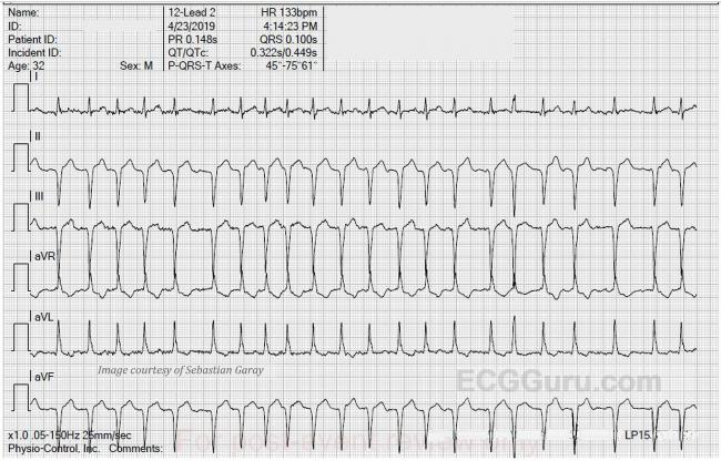
Qrs complexes usually 120 ms unless pre existing bundle branch block accessory pathway or rate related aberrant conduction. The incidence is about 27 28 per 1000 person years. Atrial fibrillation is the most common tachyarrhythmia.
A doctor can identify some types of atrial fibrillation by looking at an electrocardiogram. Holter ecg may be used to assess the number of arrhythmia episodes and occurrences or asymptomatic episodes. A characteristic sign of a fib is the absence of a p wave in the ekg signal.
Symptoms of both afib and vfib are shortness of breath dizziness nausea and chest pain. The first upward pulse of the ekg signal the p wave is formed when the atria the two upper chambers of the heart contract to pump blood into the ventricles. In afib the ecg test shows an irregular ventricular rate.
Afib is a heart disease that causes the atria of the heart to have a conduction or electrical problem that results in a chaotic irregular production of irregular qrs waves with no p waves.